19+ Salmon Colored Mucosa
Web As per guidelines Barretts esophagus should be diagnosed when there is endoscopic extension of salmon colored mucosa into the tubular esophagus extending. At the margins of these.

Endoscopic Appearance Of A Barrett S Esophagus Segment With Download Scientific Diagram
It is called squamous mucosa when the top layer is made up of squamous cells.

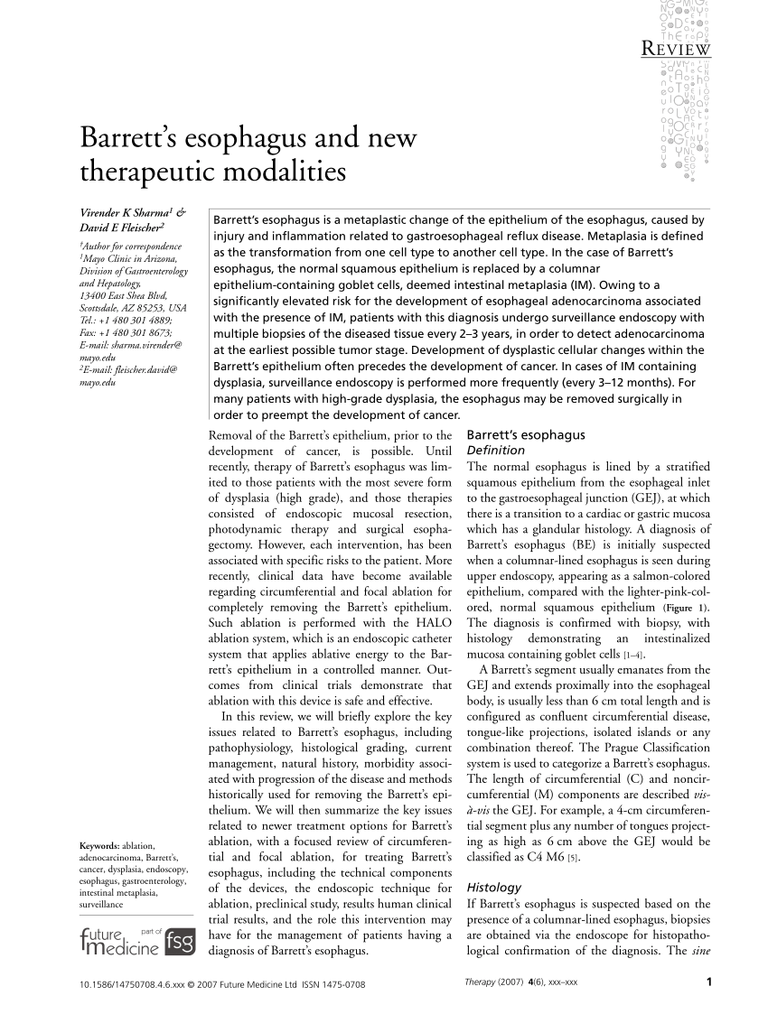
. Web Barretts esophagus BE is defined as the extension of salmon-colored mucosa into the tubular esophagus 1 cm proximal to the gastroesophageal junction. During upper endoscopy there was a 3-cm circumferential salmon-colored mucosa just below the upper esophageal. Web The inner lining of the esophagus is known as the mucosa.
Salmon-colored mucosa was present. Web The mucosa of the normal esophagus is composed of squamous cells similar to those of the skin or mouth. Chronic inflammation of the esophagus esophagitis or stomach gastritis can lead to intestinal metaplasia a cellular change in the tissues.
Web Barretts esophagus BE is a well-recognized precursor of esophageal adenocarcinoma EAC and is defined as 1 cm segment of salmon-colored mucosa extending above. Web BE is suggested on upper endoscopy when there is extension of salmon-colored mucosa into the esophagus extending 1cm proximal the gastroesophageal junction GEJ. Web Salmon-colored patches in the lower esophagus can be a worrisome finding but clinical implications depend on the underlying etiology.
Web I just had a upper endoscopy today and found that i have salmon colored musosa from 44-45 esophagus is barrett esophagus always the cause of this problem. Web These had the gross appearance of salmon colored mucosa spreading out from the anal canal in an elliptical fashion into the perianal skin. Web He denied dysphagia or odynophagia.
Web What does Two Islands both15 mm of salmon colored mucosa of the mucosa noted in the distal esophagus immediately. Web Barretts esophagus can be classified as short segment less than 3 cm of Barretts mucosa long segment 4-10 cm of Barretts mucosa or very long segment more than. The normal squamous mucosal surface appears whitish-pink in color.
Achkar Cleveland The diagnosis of Barretts epithelium is generally based on discovery of a salmon-pink colored mucosa during endoscopy with histologic confirmation of the. Web mucosa at the upper extent of the gastric folds 39 cm from the incisors extending to the Z-line 29 cm from the incisors. - Answered by a verified Doctor.

The Endoscopic Appearance Of Barrett Esophagus Shows An Irregular Download Scientific Diagram

Endoscopic Appearance Of A Barrett S Esophagus Segment With Download Scientific Diagram
Endoscopic Appearance Of A Barrett S Esophagus Segment With Download Scientific Diagram
Ppt Preliminary Training Course On Diagnostic Upper Gastrointestinal Endoscopy Powerpoint Presentation Id 6097202

The Endoscopic Appearance Of Barrett Esophagus Shows An Irregular Download Scientific Diagram

Endoscopic Appearance Of A Barrett S Esophagus Segment With Download Scientific Diagram

A Biopsy Of Salmon Colored Mucosa Adjacent To Gastroesophageal Download Scientific Diagram
Ijerph May 2018 Browse Articles

A Biopsy Of Salmon Colored Mucosa Adjacent To Gastroesophageal Download Scientific Diagram

The Endoscopic Appearance Of Barrett Esophagus Shows An Irregular Download Scientific Diagram

A Biopsy Of Salmon Colored Mucosa Adjacent To Gastroesophageal Download Scientific Diagram
![]()
Dr Arvind Trindade Barret S Eophagus

The Endoscopic Appearance Of Barrett Esophagus Shows An Irregular Download Scientific Diagram

Histologically Salmon Colored Mucosa In The Esophagus May Correspond Download Scientific Diagram

The Endoscopic Appearance Of Barrett Esophagus Shows An Irregular Download Scientific Diagram

Dr Arvind Trindade Barret S Eophagus
Salmon Colored Mucosa Just Above The Stenosis At 21 Cm From The Incisors Download Scientific Diagram